
Diagnostic hysteroscopy
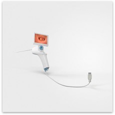
Diagnostic office hysteroscopy is a short 2-3 minutes, simple intervention that could be easily performed in the consultation room without any anesthesia with a single-use device. In more complicated cases diagnostic hysteroscopy could be also performed by a rigid optic in the operating theater under sedation. Diagnostic hysteroscopy is used to confirm the shape of the uterine cavity and excludes the presence of any anatomical abnormality or other anomalies that could affect implantation (inflammation, scar tissue).

Endometrial polyps
Endometrial polyps are a benign outgrowth of the uterine lining ranging from a few millimeters to few centimeters. They could be easily diagnosed with 2D/3D ultrasound or hysterosonography, but only hysteroscopy provides to opportunity to treat polyps by removing and analyzing them. Smaller polyps (less than 10 mm) could be monitored to see if they disappear spontaneously, but larger ones need to be removed as they could interfere with embryo implantation or an early pregnancy.
Submucosal fibroids
Uterine fibroids are common benign tumors in women of reproductive age arising from the muscle tissue of the uterus. Depending on their position those fibroids that encroach on the uterine cavity (intracavitary, submucosal or submucosal/intramural) should be removed as they could cause significant symptoms and interfere with embryo implantation and early pregnancy. A diagnostic hysteroscopy allows direct visualization of the fibroid and planning for an operative intervention.
Intrauterine adhesions
Intrauterine adhesions or Asherman syndrome is caused by trauma to the inner layer of the endometrial lining usually after surgical intervention on a gravid uterus (after a miscarriage or complicated delivery). A diagnostic hysteroscopy could provide an initial diagnosis and the extent of intrauterine adhesions and allows for planning of an operative intervention (sometimes repeated) in the hand of skilled endoscopic surgeon.
Uterine malformations
The incidence of congenital uterine abnormalities could be as high as 8% in infertile women. These could be diagnosed less invasively by 3D ultrasound and hysterosonography, but a diagnostic hysteroscopy allows a direct visualization of the uterine cavity. Some uterine malformations (septate uterus, dysmorphic uterus) could be managed surgically with an operative hysteroscopy in hands of a well-trained gynecologic surgeon.
Other abnormalities like chronic endometritis (inflammation) or signs of uterine adenomyosis could also be diagnosed by a diagnostic hysteroscopy. The confirmation of a completely normal uterine cavity in patients with recurrent implantation failure indicates that other investigations need to be conducted to search for other potential causes of failed implantation.